

Histology
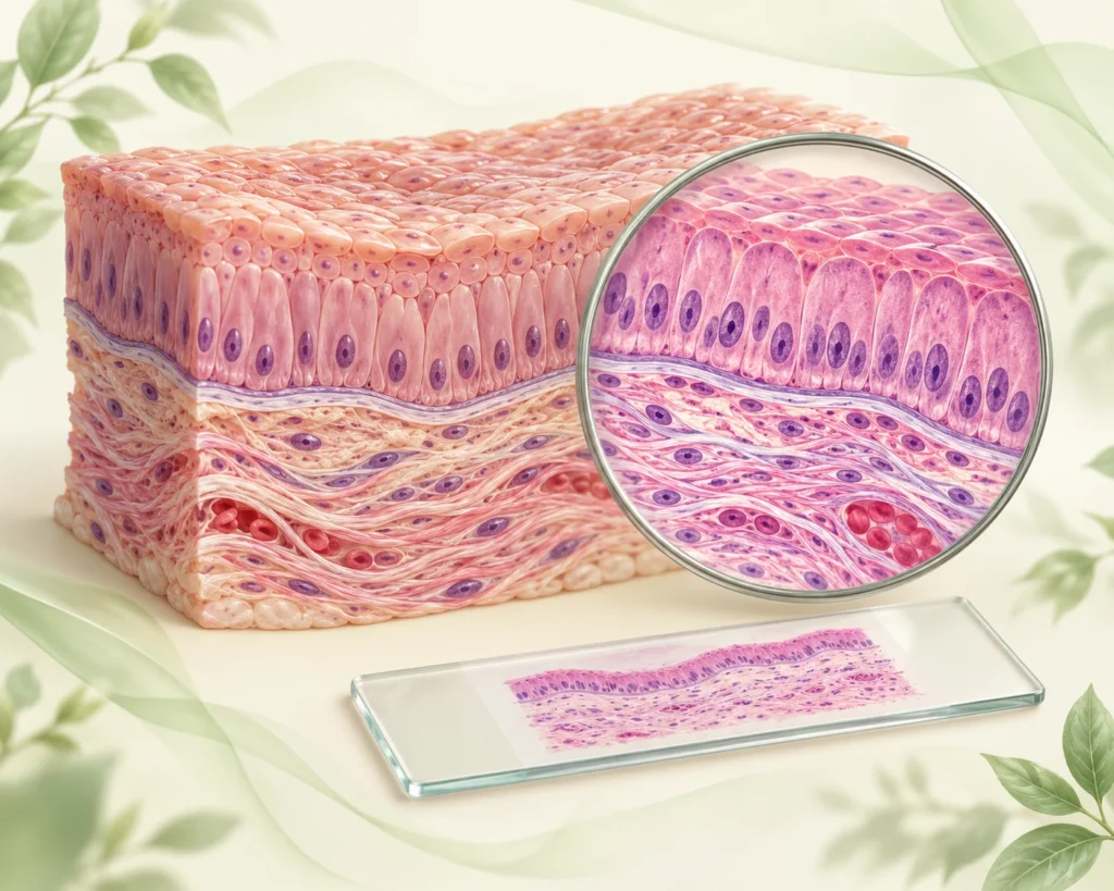
Histology is the microscopic study of cells, tissues and organs and explains how microscopic structure supports normal function.
General subject summary is free to everyone.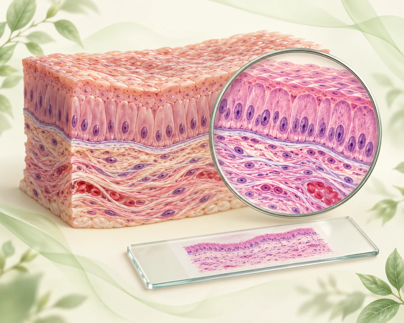
Histology
Subject Summary
The public overview below follows the supplied PhysioVeda Academics subject summary.
Histology is the microscopic study of cells, tissues and organs and explains how microscopic structure supports normal function.
A practical approach is to identify the dominant cell type, extracellular matrix, tissue organization, surface specialization and vascular or neural relationships seen in each specimen.
Basic tissues - epithelium, connective tissue, muscle and nervous tissue - combine in characteristic patterns to form organs, while specialized histology explains filtration, secretion, absorption, conduction, contraction and sensory transduction.
For medical and rehabilitation sciences, histology provides the structural basis for understanding tissue healing, degeneration, fibrosis, muscle and nerve injury, bone remodeling and organ disease.
This summary follows the uploaded PhysioVeda Academics structure from microscopy and basic tissues through organ-system histology and the special senses.
Whole-subject high-yield numbers
Use these supplied values and key facts for quick whole-subject revision.
| Four basic tissue classes | Epithelium, connective tissue, muscle, nervous tissue |
| Light microscopy | Uses visible light; routine paraffin sections commonly stained with H&E |
| Electron microscopy | Transmission EM shows internal ultrastructure; scanning EM shows surface topography |
| Epithelium | Polarized cells with little extracellular matrix and basement-membrane attachment |
| Cartilage | Avascular; chondrocytes in lacunae |
| Bone | Mineralized type I collagen matrix with continuous remodeling |
| Skeletal muscle | Striated, multinucleated, voluntary |
| Cardiac muscle | Striated, branched, intercalated discs |
| Smooth muscle | Non-striated, spindle-shaped, involuntary |
| Blood-air barrier | Type I pneumocyte + fused basal laminae + capillary endothelium |
| Renal filtration barrier | Fenestrated endothelium + glomerular basement membrane + podocyte slit diaphragm |
Core references
- Mescher AL. Junqueira's Basic Histology: Text and Atlas. 17th ed. McGraw Hill.
- Pawlina W. Histology: A Text and Atlas. 9th ed. Wolters Kluwer.
- Gartner LP, Hiatt JL. Color Textbook of Histology. 5th ed. Elsevier.
Want to go deeper into Histology?
Continue into the complete topic and subtopic learning system with structured summaries, medical visuals, topic wise OBQ practice, timed tests, explanations and results.